Every cell you have holds a voltage across its outer membrane, and it spends about a fifth of your food to keep it there. The table below is the whole subject in one screen: what the number is for each kind of cell, and what happens when it moves.
| Cell or state | Resting voltage | What that voltage goes with | Measured in | Source |
|---|---|---|---|---|
| Neuron, skeletal muscle | −70 to −90 mV | Fully differentiated, not dividing | Human and animal tissue | PMID 23882223 |
| Stem and progenitor cells | about −20 to −40 mV | Still dividing, identity not yet fixed | Human mesenchymal stem cells | PMID 19011685 |
| Tumour cell | −10 to −40 mV | Dividing; the voltage shifts before the tissue looks abnormal | Frog embryo, human cell lines | PMID 23882223, PMID 23471912 |
| Frog tail cell pushed more negative with a K⁺ channel | more negative than its neighbours | Builds a complete eye, with lens, retina and optic nerve | Frog (Xenopus) embryo | PMID 22159581 |
| Planarian head wound with its H⁺/K⁺ pump blocked | held at the wrong value for a head | Builds a tail where a head belongs | Planarian flatworm | PMID 21276941 |
| Any wound edge | 100 to 300 mV per millimetre of field | Cells crawl along the field toward the damage | Multiple species, including human cells | PMID 15987799 |
The first three rows are the pattern that makes this subject interesting rather than a footnote in a physiology textbook: voltage tracks what a cell is doing, not just whether it can fire. A neuron at −80 mV is a cell that has stopped dividing and settled into a job; a tumour cell at −20 mV is a cell that has not. The last three rows are the harder claim, that moving the voltage on purpose changes what a tissue builds. Every one of them is a published animal result with a PMID beside it, and none of them is a treatment for anything.
What the number is actually measuring
Membrane potential is a difference in electric charge between the inside of a cell and the fluid around it, held in place by a difference in ion concentrations. Three things produce it and all three are running at once.
Pumps spend energy to move ions the wrong way. The sodium-potassium pump pushes three sodium ions out for every two potassium ions it brings in, and it pays one ATP for each cycle. It runs against the concentration gradient, which is the point: it is what builds the gradient.
Channels let ions leak back. A resting cell is much more permeable to potassium than to anything else. Potassium leaves down its concentration gradient, and because it carries a positive charge with it, the inside is left more negative than the outside.
The charge separation stops the leak. Potassium keeps leaving until the growing negative charge inside pulls it back as fast as the concentration difference pushes it out. That balance point is a voltage, and it is calculable.
🛑 This is not electricity in the wall-socket sense, and the difference is not pedantry. Nothing here is a current running down a wire, nothing arcs, nothing is felt, and nothing a person does with a device at home changes it. It is a difference in the number of charged atoms on two sides of a four-nanometre layer of fat, maintained by protein machines, and it is read out with dyes and glass electrodes in a laboratory.
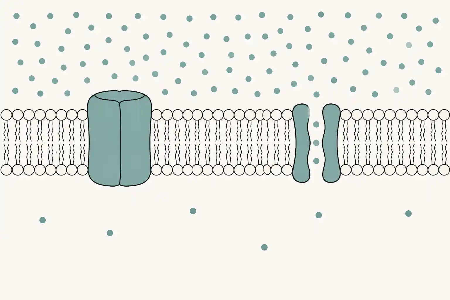
The picture is here for one reason the prose cannot do as quickly: the voltage exists ACROSS a surface, not inside a volume. People new to the subject picture a cell as a battery with charge stored in the middle, and then every later claim reads wrongly. The charge is a thin excess on each face of the membrane, held apart by the membrane itself; the bulk of the cytoplasm is electrically neutral. The proteins spanning the layer are the pumps that build the gradient and the channels that let it leak back, and the whole balance between those two is what the number in the previous table reports.
Work the number out yourself
For one ion at a time the balance point is the Nernst potential, and at body temperature it is one multiplication. The constant is 61.5 mV per tenfold concentration difference at 37 °C, for an ion carrying one positive charge.
E = 61.5 × log₁₀(concentration outside ÷ concentration inside)
Take potassium, with the standard textbook values of about 140 mM inside a cell and 5 mM in the fluid around it:
E = 61.5 × log₁₀(5 ÷ 140)
E = 61.5 × log₁₀(0.0357)
E = 61.5 × (−1.447)
E = −89 mV
That is within a few millivolts of the resting potential measured in a real skeletal muscle fibre, which is the reason potassium is described as the ion that sets the resting potential. Run the same line for sodium, at about 12 mM inside and 145 mM outside, and it comes out at +67 mV — the opposite sign, and nowhere near the resting value, because a resting membrane barely lets sodium through at all. The real resting potential sits close to whichever ion the membrane is most permeable to, and that is what makes permeability, rather than concentration, the thing a cell changes when it wants to move its voltage.
If X then Y
- If a cell opens more potassium channels, it moves toward −89 mV and gets more negative. That is hyperpolarisation.
- If a cell opens sodium or chloride channels, it moves away from −89 mV and gets less negative. That is depolarisation.
- If the pump stops, the gradients run down over minutes to hours rather than instantly, because the gradient is a store the leak has to drain.
Why holding it costs a fifth of your food
The sodium-potassium pump never stops. Across the whole body it runs at roughly 20 to 25 per cent of all the ATP produced, and in the brain at 60 to 70 per cent (PMID 18128147). A cell pays that bill every second it is alive, to hold a number around −70 mV.
That price is the argument that the voltage is doing more than keeping nerves excitable, because most cells in your body never fire anything. Skin, liver, bone and gut cells all hold a resting potential and none of them sends an action potential. Something is being paid for.
This is the single most useful thing to know before reading anything else in this field, and it is the field's own standard (PMID 26864374). Knocking down an ion channel changes two things at the same time: a protein disappears, and the cell's voltage moves. Any result from that experiment alone is ambiguous, because the protein may have a job that has nothing to do with charge. The test that separates them is heterologous rescue — put the voltage back using a completely unrelated channel, one that shares no sequence and no binding partners with the first, and see whether the defect is repaired. In the craniofacial work that established the method, it was. Many published results in this area do not meet that bar, and reading the literature with that one filter switched on is the honest way to do it.
| What was done | To what | What happened | Why it is hard to explain another way | Source |
|---|---|---|---|---|
| Blocked the H⁺/K⁺ pump with SCH-28080 at a head wound | Planarian flatworm | The fragment regrew a tail where a head belonged | An untreated fragment cut anywhere rebuilds the right end; the identity of the part is decided at the wound | PMID 21276941 |
| Expressed the Kir2.1 potassium channel to hyperpolarise cells | Frog embryo gut, tail and somite | A complete eye formed, with lens, retina and optic nerve | It only works in tissue that is already competent, so the voltage is an instruction about WHICH organ, not a printer that builds one | PMID 22159581 |
| Trained tadpoles carrying an eye on the tail | Frog tadpole | They learned visual tasks through it | Nothing in the animal was told to expect an eye there, and the nervous system used it anyway | PMID 23447666 |
| Briefly blocked gap junctions, then cut the worms again in plain water | Planarian flatworm | Two heads again, round after round, with the genome untouched | The pattern survives in something that is not the DNA sequence; nobody has identified where | PMID 28538159 |
| Blocked the same pump with omeprazole in a 4-cell embryo | Frog embryo | Which side the heart formed on was randomised | The step happens before the cilia textbooks credit with left-right asymmetry | PMID 12372302 |
Read this table as a list of decisions a tissue makes, not as a list of things voltage can build. In every row the experiment moved a voltage and the tissue built a different but perfectly ordinary structure — a tail, an eye, a second head, a mirrored body axis. Nothing was assembled cell by cell and no shape was specified in detail. That distinction is the whole difference between what has been demonstrated and the thing the field is aiming at, and the honest status of the aim is that it does not exist yet (PMID 33826908). Every row is an animal result. None of it is a therapy, and nothing on this page is medical advice.
What goes wrong, and what it costs
There is no instrument that reads voltage inside a living human. The measurements above come from glass microelectrodes in single cells and from voltage-sensitive dyes in transparent embryos (PMID 22474652). Neither works through skin. Any claim about a person's own tissue voltage, measured by anything a consumer can buy, is describing something that has not been measured.
A dye is not a voltmeter. Voltage-sensitive dyes such as DiBAC report a relative brightness, not a number in millivolts, and they respond to other things — membrane binding, dye loading, cell volume. The frog-embryo result where a dye picked out a pre-tumour patch before any microscope could see one (PMID 23471912) is a comparison between neighbouring cells in the same image, not an absolute reading.
Correlation is the default state of this literature. Tumour cells sitting depolarised is one of the best established facts in the area and it still does not say which way the arrow runs. Whether voltage is a cause, a consequence, or both at different stages of the same disease is unresolved, and no human therapy exists (PMID 23882223).
The word "bioelectricity" invites the wrong picture, and that is a cost. It is the pattern of ion concentrations and voltages across tissue. It is not energy, not frequency, not vibration, and not anything a person can do to themselves. The signalling layer is ancient enough that bacteria in a biofilm coordinate with propagating potassium waves through the same kind of channel (PMID 26503040), which is a much stranger fact than any of the ones people invent.
Key Takeaways
- Membrane potential is the voltage across a cell's outer membrane, around −70 to −90 mV in a differentiated human cell and −10 to −40 mV in a tumour cell (PMID 23882223).
- It is built by pumps spending ATP and set by whichever ion the membrane currently lets through most easily.
- One multiplication gives the potassium balance point: 61.5 × log₁₀(5 ÷ 140) = −89 mV at 37 °C, which is close to a real muscle fibre's resting value.
- Holding it costs roughly 20 to 25 per cent of whole-body ATP and 60 to 70 per cent of the brain's (PMID 18128147).
- Moving it on purpose changes what a tissue builds: a tail instead of a head in a planarian (PMID 21276941), a working eye on a tadpole's tail (PMID 22159581, PMID 23447666).
- A knockdown alone proves nothing. The test is heterologous rescue: put the voltage back with an unrelated channel and see if the defect goes (PMID 26864374).
- No instrument reads voltage inside a living human (PMID 22474652), and nothing on this page is a treatment.